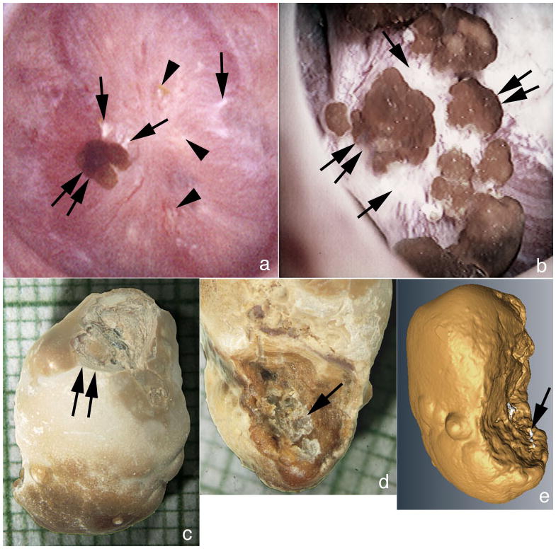
Figure 2. Details of attached stones.

(a) An attached stone (double arrow) is seen resting on a region of white plaque (single arrows) and intermixed with small areas of white (single arrow) and yellow plaque (arrowheads). Patient 8 had numerous attached stones (double arrow) atop an extensive area of white plaque (single arrows) much like that found in some ICSF. Analysis of attached stones by μ-CT revealed these to be composed of primarily of CaOx with small sites of apatite corresponding to a site of attachment to white (Randall’s) plaque. (c & d) g Light microscopic image of an attached revealing the smooth urinary (c) and papillary (d) surface morphology. The papillary surface (d) shows a concave region with crystalline material (single arrow) consistent with an attachment site. The urinary surface (c) shows a damaged region (double arrow) generated during stone removal. (e) Reconstruction of μ-CT images shows regions of CaOx in yellow and areas of apatite in white. The white regions are appear to present the attachment site.
