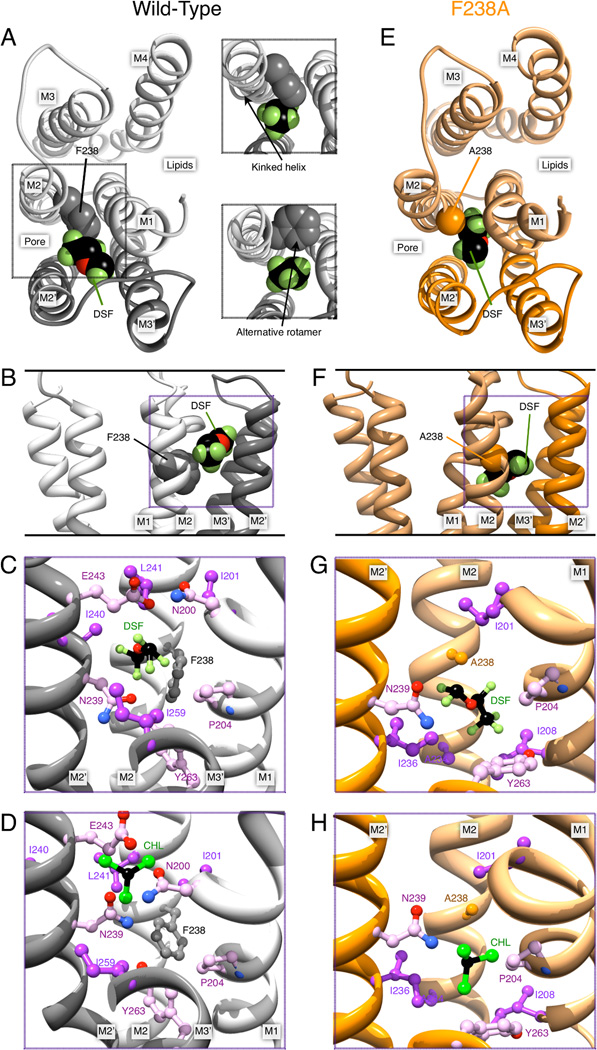
Figure 4.
Desflurane (DSF) and chloroform (CHL) poses in the inter-subunit cavity of GLIC. (A) Left, GLIC structure with desflurane in the inter-subunit binding site, viewed from the extracellular side. For clarity, only M1–M4 of the upper subunit (light gray) and M2–M3 from the lower subunit (dark gray) are shown. F238 and helices contributing to the intra-subunit cavity are labeled. Right, forcing the ligand to bind in the WT close to F238 either induces a kink in M2, or requires F238 to adopt a different rotamer. (B) Desflurane binding viewed in the plane of the membrane in the pore. (C) Inter-subunit environment for desflurane binding in WT GLIC; note the close proximity to F238. (D) Chloroform bound to the inter-subunit site of WT GLIC. Panels (E) through (H) contain corresponding illustrations for ligands bound to GLIC F238A. The mutation yields a much larger cavity for ligand binding between the subunits. In particular, in panels (G) and (H), desflurane and chloroform assume poses that would directly overlap with the F238 side chain.

