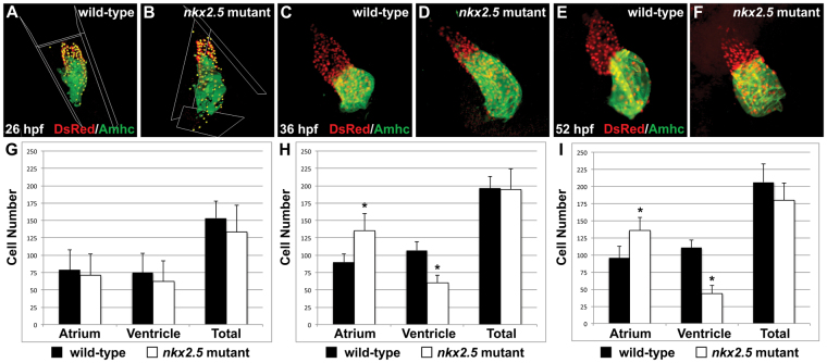
Fig. 2.
Increased atrial and decreased ventricular cell numbers are first evident in nkx2.5 mutants following heart tube extension. (A-F) Immunofluorescence indicates expression of the transgene Tg(-5.1myl7:nDsRed2) (red) in both cardiac chambers facilitating cardiomyocyte counting at 26 hpf (A,B), 36 hpf (C,D) and 52 hpf (E,F). Atria are labeled with the anti-Amhc antibody S46 (green). (G-I) Bar graphs indicate numbers of atrial and ventricular cardiomyocyte nuclei, as well as the total number of cardiomyocytes; mean and s.e.m. of each data set are shown, and asterisks indicate statistically significant differences from wild type (P<0.001). (G) At 26 hpf, we find no statistically significant difference in cell numbers in wild-type (n=20) and nkx2.5 mutant (n=7) embryos. (H) At 36 hpf, comparison of wild-type (n=16) and nkx2.5 mutant (n=10) embryos reveals an increase in atrial cell number and a decrease in ventricular cell number in nkx2.5 mutants. (I) At 52 hpf, comparison of wild-type (n=16) and nkx2.5 mutant (n=11) embryos reveals an increase in atrial cell number and a decrease in ventricular cell number in nkx2.5 mutants.

