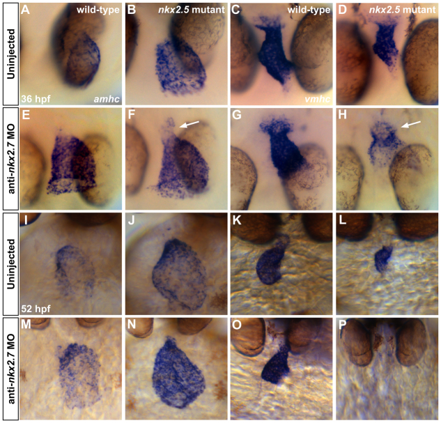
Fig. 6.
Gradual elimination of ventricular gene expression and expansion of atrial gene expression in the Nkx-deficient heart. In situ hybridization illustrates expression of amhc (A,B,E,F,I,J,M,N) and vmhc (C,D,G,H,K,L,O,P) in wild-type embryos (A,C,I,K), nkx2.5 mutants (B,D,J,L), wild-type embryos injected with anti-nkx2.7 MO (E,G,M,O) and Nkx-deficient embryos (F,H,N,P). (A-H) Dorsal views, anterior to the bottom, at 36 hpf. The nkx2.5 mutants display a dilated atrium (A,B) and shortened ventricle (C,D). Following MO injection, wild-type embryos demonstrate a swollen atrium and widened ventricle (E,G). In nkx2.5 mutants, MO injection leads to distinct expansion of amhc and fading of vmhc in the ventricular remnant (F,H; arrows). (I-P) Ventral views, anterior to the top, at 52 hpf. Phenotypes are exacerbated at 52 hpf, both in terms of morphological defects and expression pattern changes. Most dramatically, in the Nkx-deficient embryo, amhc is expressed throughout the entire heart whereas vmhc expression is effectively abolished (N,P).

