
Fig. 1.
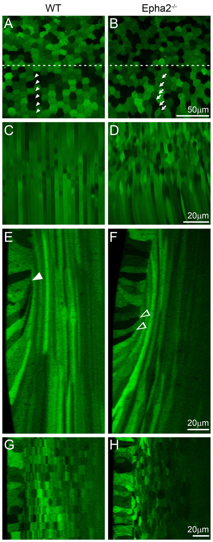
Confocal images of the equatorial region of P21 GFP+ wild-type and Epha2-/- mouse lenses. (A-D) The wild-type (WT) lens shows the typical mosaic GFP expression pattern in the equatorial epithelial cells (A), with hexagonal cells organized into meridional rows (below the dashed line, arrowheads) and straight and organized peripheral differentiating fiber cells (C). By contrast, equatorial epithelial cells in the Epha2-/- lens lack organized meridional rows (B, arrows), and underlying fiber cells are wavy and disorganized (D). (E,F) Three-dimensional reconstruction of z-stacks in the anterior-posterior view through the WT lens reveals organized fiber cells in the bow region (E), whereas a comparable reconstruction through the Epha2-/- lens demonstrates disorganization of peripheral fiber cells with variable cell width (F). The WT lens fulcrum is indicated by the arrowhead (E); in the Epha2-/- lens, multiple points of apical constriction are observed (F, arrowheads). (G,H) Reconstruction of z-stacks through the transverse view shows that WT fiber cells are neatly organized into straight rows (G), whereas Epha2-/- fiber cells are misaligned (H). The epithelial cells are on the left in G and H.