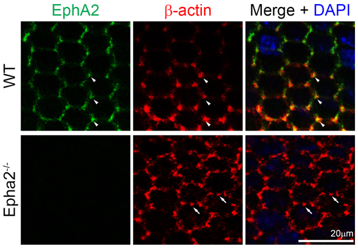
Fig. 2.

EphA2 and β-actin distribution in WT and Epha2-/- equatorial epithelial cells. Double immunolabeling of β-actin (red) and EphA2 (green) with DAPI staining (blue, nuclei) of equatorial lens epithelial cells from lens capsule flat-mounts of P21 WT and Epha2-/- mice. When equatorial cells organize into meridional rows, β-actin and EphA2 are enriched and colocalize at the vertices of hexagonal WT epithelial cells (arrowheads). By contrast, β-actin forms abnormal aggregates on the membranes of Epha2-/- epithelial cells that fail to pack into organized meridional rows (arrows). Antibody specificity is demonstrated by the lack of EphA2 staining in Epha2-/- cells.
