Fig. 4.
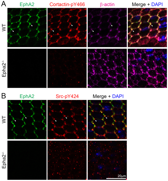
EphA2, phosphorylated cortactin, β-actin and phosphorylated Src distribution in WT and Epha2-/- equatorial epithelial cells. (A) Triple immunolabeling of EphA2 (green), phosphorylated cortactin-Y466 (cortactin-pY466, red) and β-actin (purple) with DAPI staining (blue, nuclei) of equatorial lens epithelial cells from lens capsule flat-mounts of P21 WT and Epha2-/- mice. In hexagonal WT equatorial epithelial cells, EphA2, cortactin-pY466 and β-actin are localized at the cell membrane and are enriched and colocalize at cell vertices (arrowheads). By contrast, only punctate staining signals for cortactin-pY466 can be found in the disorganized Epha2-/- epithelial cells. (B) Similarly, double immunolabeling of phosphorylated Src-Y424 (Src-pY424, red) and EphA2 (green) reveals that both proteins are enriched and colocalize at the vertices of hexagonal WT epithelial cells (arrowheads). However, in the disorganized Epha2-/- epithelial cells, only random punctate signals from Src-pY424 are observed.

