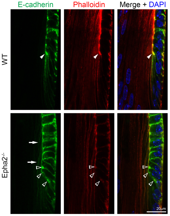
Fig. 6.

E-cadherin and phalloidin staining of P14 WT and Epha2-/- frozen lens sections. Double immunolabeling of E-cadherin (green) and with phalloidin (F-actin, red) in frozen lens sections demonstrates that E-cadherin and F-actin are highly enriched only at the lens fulcrum (arrowheads) in the WT lens section, but form multiple aggregates in the Epha2-/- lens section (open arrowheads). Abnormal E-cadherin signal is observed in Epha2-/- lens fiber cells (arrows).
