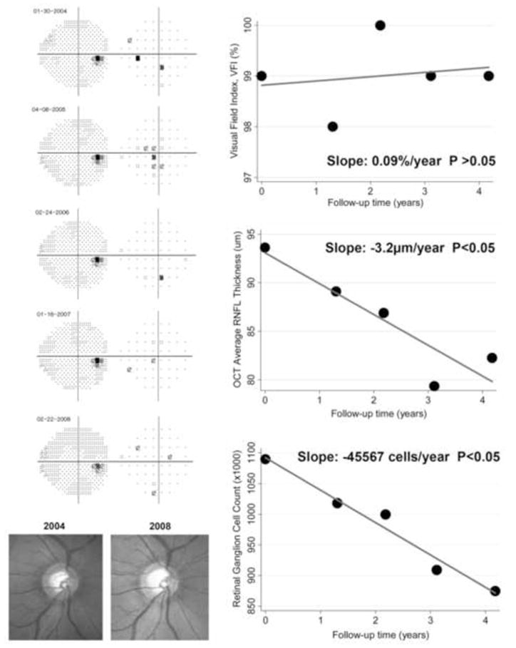
Figure 5.

Eye detected as progressing by the rate of retinal ganglion cell loss with a slope of −45567 cells/year (P<0.05), but not by the Visual Field Index. The eye had early glaucomatous damage and showed progressive neuroretinal rim thinning as seen on the optic disc stereophotographs. The optical coherence tomography parameter average thickness showed a statistically significant slope of −3.2μm/year.
