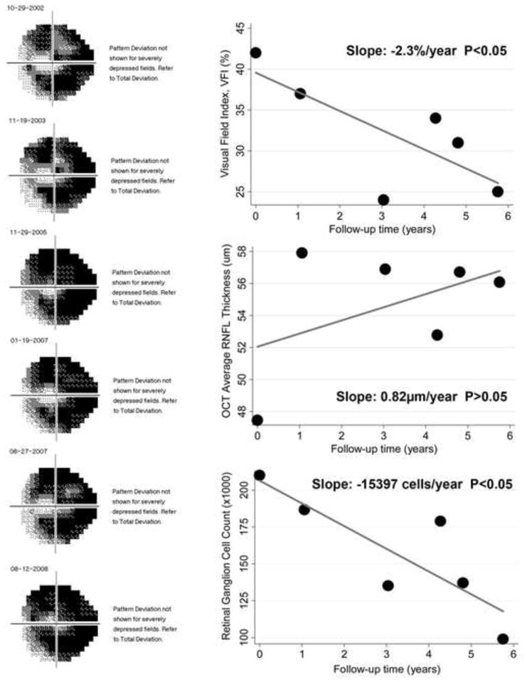
Figure 6.

Eye detected as progressing by the rate of retinal ganglion cell (RGC) loss with a slope of −15397 cells/year (P<0.05), but not by the optical coherence tomography average thickness parameter. The eye had advanced visual field loss and a statistically significant slope of change with the Visual Field Index (−2.3%/year).
