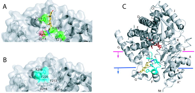
Figure 3.

Peripheral ligand binding site in CYP3A4. A, A progesterone binding site identified in the 1WOF crystal structure.17 The progesterone molecule (shown in yellow) is located above the Phe-cluster (side chains are displayed) and forms a hydrogen bond with the amide nitrogen of Asp214 (depicted as a dotted line) and hydrophobic interactions with Leu211, Phe213 and Phe219 (shown in green). B, A high affinity binding site for Fluorol-7GA identified with FRET34 is located on the surface near residues 217-220 (shown in cyan) and overlaps with the progesterone binding site. C, A cartoon diagram of CYP3A4 with two possible levels of incorporation into the membrane bilayer.35,36 The heme is shown in red sticks, progesterone in yellow, and the progesterone/Fluorol-7GA binding site in cyan.
