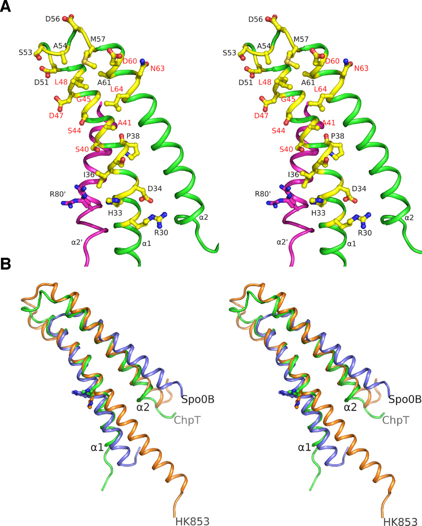
FIGURE 3.
Stereoview of the ChpT recognition surface for RRs. A. Residues (shown in yellow/blue/red ball-and-sticks) in the ChpT DHp region (green) hypothesized to be involved in binding RRs or phosphotransfer. The residues mutated in this study are labeled in red colored texts. B. Structural alignments of the DHp regions of ChpT (green), HK853 (orange) and Spo0B (blue). ChpT has a shorter α1 helix like Spo0B compared to HK853. Histidine residues involved in phosphotransfer are shown as balls-and-sticks (see also Fig. S2).

