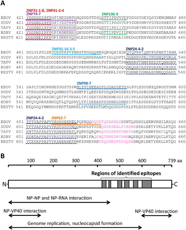
Fig. 3.
Epitope sequences in amino acid sequence alignment and known functional regions NPs. (A) Amino acid sequences of EBOV, SUDV, TAFV, BDBV and RESTV were obtained from GenBank under accession numbers AF272001, AF173836, FJ217162, FJ217161 and AF522874, respectively. Amino acid sequences at positions 421–660 of each virus are shown. EBOV NP peptides recognised by the mAbs are highlighted with solid lines. Corresponding regions of the other NPs to which each mAb showed strong cross-reactivity are underlined (dashed lines). Amino acid sequences used for producing species-specific rabbit antisera are shown in pink. (B) Locations of the identified epitopes are shown in the schematic diagram of NP. Functional domains (Bharat et al., 2012; Noda et al., 2007; Watanabe et al., 2006) are also shown. (For interpretation of the references to colour in this figure legend, the reader is referred to the web version of the article.)

