
Fig. 1.
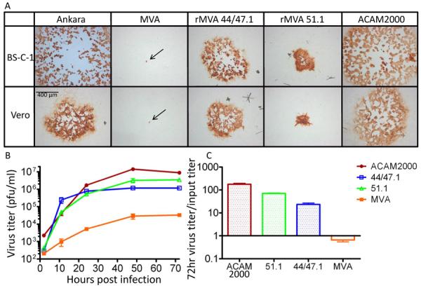
Replication of rMVAs. (A) Cell-to-cell virus spread. Monolayer of BS-C-1 and Vero cells were infected with MVA, rMVA 44/47.1 and rMVA 51.1, Ankara and ACAM2000 viruses. At 48 h after infection, the cells were fixed and immunostained with broadly reactive anti-VACV antibody followed by horseradish peroxidase conjugated to anti-rabbit immunoglobulin. Arrows point to the pin-point foci formed by MVA. (B) Growth curve. Vero cells were infected with 0.1 pfu/cell of MVA, rMVA 44/47.1, rMVA 51.1 or ACAM2000 and harvested at the indicated times post infection. Cell-associated viruses were titrated on CEF monolayers in the case of MVA and rMVAs and BS-C-1 cells for ACAM2000. Standard error bars shown. (C) The ratios of virus output to virus input are plotted with standard error bars.