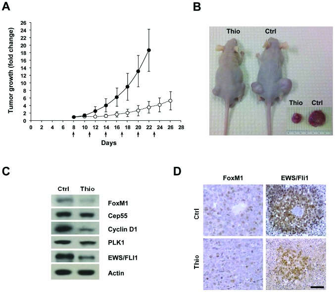
Figure 4.
Thiostrepton treatment reduces the tumorigenicity of EWS cells. (A) Xenografts established by s.c. injection of A4573 cells in athymic (BALB/c nu/nu) nude mice were given i.p. dosage of thiostrepton (17 mg/kg) (n=10). Tumor growth in the animals was monitored on alternate days following s.c. cell injections, and statistically significant differences in tumor volume were noted for the treated sample around 22 days post-injection (p≤0.00002). Arrows indicate days when the mice were subjected to thiostrepton injection. (B) Mice injected with A4573 cells and treated with vehicle or with thiostrepton were euthanized when tumors in control animals reached maximum allowable tumor burden (22 days), and tumors were excised and their volume measured. Inset shows images of representative tumors isolated at 22 days post s.c. cell injection. (C) Immunoblot showing the reduction in the levels of FoxM1 and some of its transcriptional targets as well as of the EWS/FLI1 oncoprotein in lysates from mice treated with thiostrepton relative to those from untreated animals; β-actin was used as loading control. (D) Sections of xenograft tumors excised from mice treated with vehicle or triostrepton were subjected to immunohistochemical analysis to detect FoxM1 or EWS/FLI1. Results showed decreased staining for both target proteins in tumors excised from thiostrepton-treated mice relative to those obtained from control animals. Scale bar, 50 μm. Ctrl and Thio represent control and thiostrepton-treated samples.

