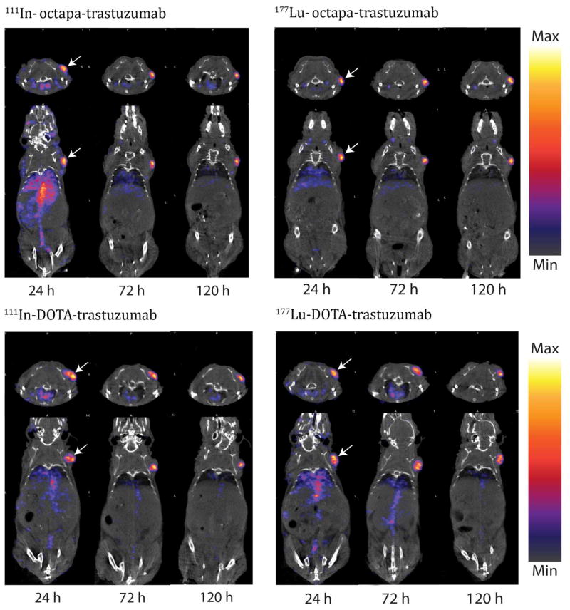
Figure 2. SPECT/CT imaging of the 111In/177Lu-octapa-trastuzumab and 111In/177Lu- DOTA-trastuzumab immunoconjugates.
Imaging studies in female nude athymic mice with subcutaneous SKOV-3 xenografts (identified by arrow at right shoulder, tumor volume ~ 100–150 mm3), showing transverse (top) and coronal (bottom) planar images bisecting the tumor, imaged at 24, 48, 72, 96, and 120 h post injection, see Figures S17–20 for 48 and 96 h time points.

