
Fig. 4.
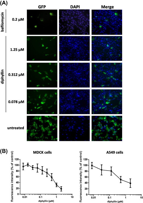
Diphyllin inhibited the GFP expression from the NS1-GFP influenza virus. 0.2 μM of bafilomycin A1 or various concentrations of diphyllin (0.078, 0.312, 1.25 μM) were added to MDCK cells 1 h before NS1-GFP virus infection (MOI = 0.01). Infected cells without diphyllin treatment were used as controls. After a 1-h period of infection, cells were washed, overlaid with fresh media containing the same concentrations of diphyllin as in previous step, and incubated for another 24 h. (A) Fluorescence images of GFP (green) and nucleus (DAPI, blue) were acquired using DeltaVision deconvolution microscope system. Representative images are shown (magnification: 200×). (B) Green fluorescence intensity from diphyllin-treated cells was quantitated using an iCys Research Imaging Cytometer. Data was presented by the relative intensity of untreated controls cells. Values are mean ± SD from four replicates. (For interpretation of the references to color in this figure legend, the reader is referred to the web version of this article).