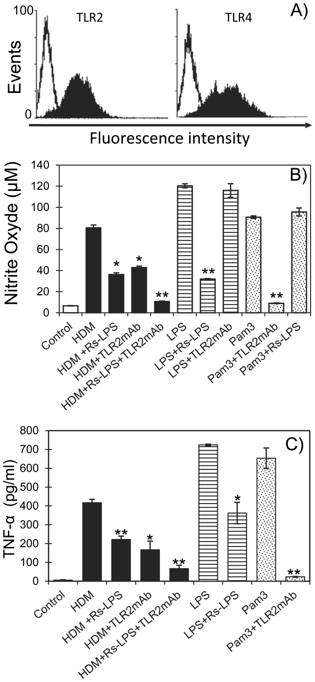
Figure 3. TLR dependent NO and TNF-α production by HDM stimulated MH-S cells.
(A) Evidence of constitutive expression of TLR2 and TLR4 in MH-S cells. Cells were incubated with mouse anti-TLR2 and -TLR4 antibodies and analyzed by flow cytometry. Effects of TLR2 and TLR4 antagonists on the HDM extract-induced NO (B) and TNF-α (C) production by MH-S cells. MH-S cells were stimulated by the HDM extract (10 µg/mL), E. coli LPS (0.1µg/mL) or Pam3CSK4 (0.01µg/mL) in absence or presence of anti-TLR2 monoclonal antibodies (TLR2mAb) or/and of the TLR4 antagonist LPS from Rhodobacter sphaeroides (Rs-LPS, 10µg/mL). After 48h supernatants were collected and assayed for NO and TNF-α production as described in Material and Methods. All samples were analyzed in triplicate and bars represent the mean of the relative values ± SEM from two independent experiments. Statistical analysis was performed using Student’s t-test; *p<0.05, **p<0.01.

