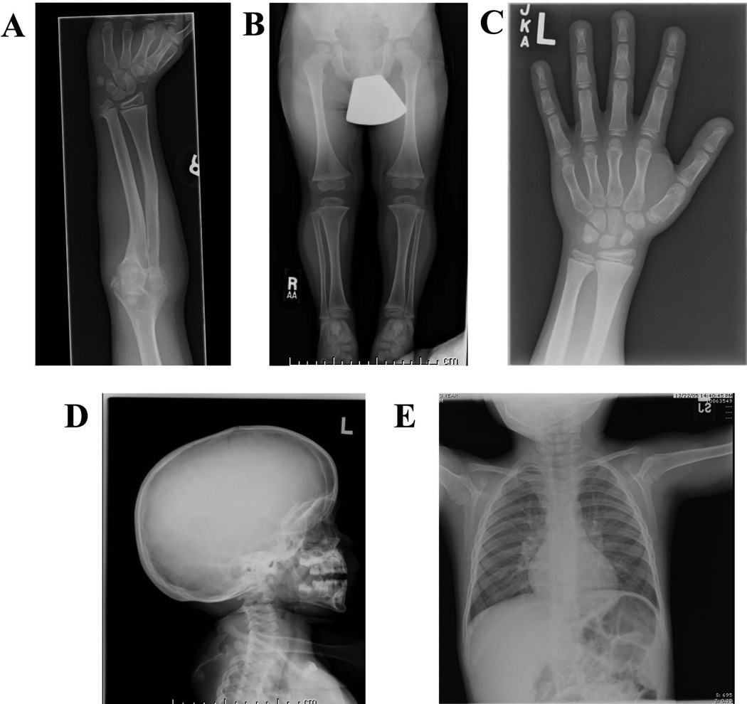
Figure 2.
Radiographs highlighting various skeletal features in the patient. Radiographs were taken from various skeletal surveys conducted at different ages. A) Ulnar bowing of radial shaft and radioulnar synostoses (9 years old). B) Flaring of proximal tibial metaphyses (3 years old). C) Bulbing of phalangeal tufts (8 years old). D) Dolichocephaly (3 years old). D) Chest x-ray showing normal clavicles (3 years old).

