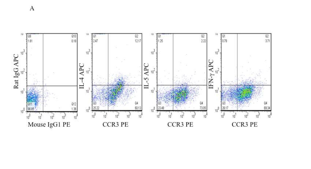
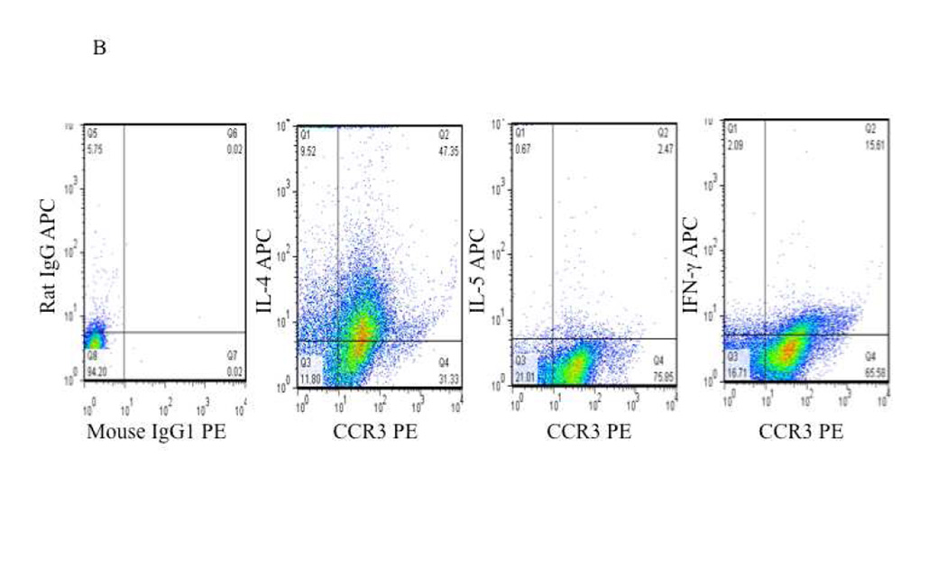
Figure 2. Intracellular cytokine staining of CCR3 positive eosinophils in CHES and AERD.
Nasal polyps were digested and cells were separated from collagenous support material. Cells were stained with appropriate markers and analyzed by flow cytometry. Initial gating was performed to include only CD45+ cells. This was followed by extracellular staining for CCR3, CD4, CD8, or CD19 and intracellular staining for cytokines. A. Representative staining of CHES nasal polyp cells that are CCR3+ showing intracellular levels of IL-4, IL-5 and IFN-γ. B. Representative staining of AERD nasal polyp cells that are CCR3+ showing intracellular levels of IL-4, IL-5 and IFN-γ.


