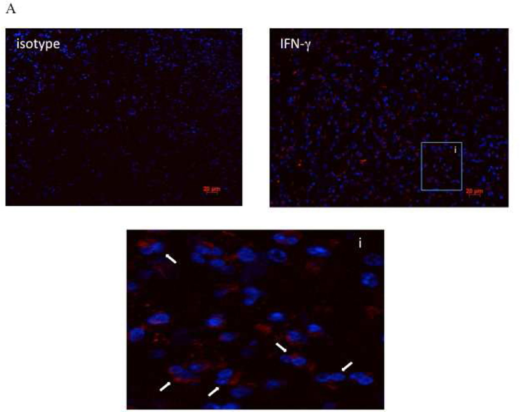
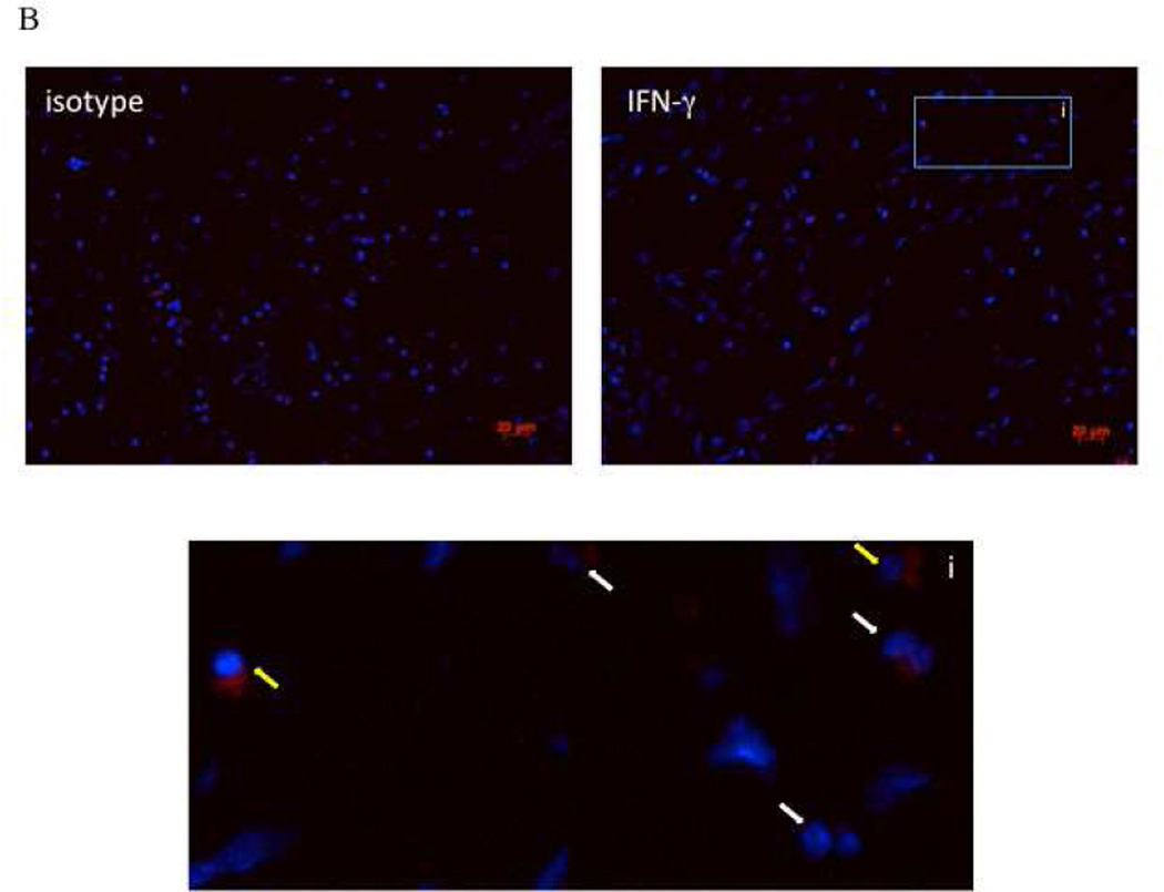
Figure 3. Immunofluorescence for IFN-γ in AERD and CHES polyps.
Sections from paraffin-embedded nasal polyps from AERD (3A) and CHES (3B) subjects were stained with an APC-labeled antibody directed against IFN-γ and counterstained with DAPI to display nuclei. Each grouping displays an isotype control, specific IFN-γ staining and an enlarged region to show specific cells. In these figures, red represents IFN-γ staining, blue is nuclear staining, i=insert, white arrows show eosinophils, and yellow arrows show mononuclear cells.


