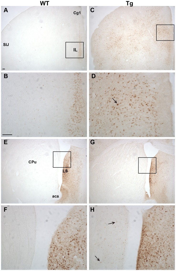
Figure 3. Ngb immunoreactivity (IR) in the forebrain.
WT Ngb-IR was mostly confined to a thin layer of the Il and Cg1 (A-B). In Tg mice Ngb-IR was seen throughout the forebrain in medium intense stained cells (C-D). Intense Ngb-IR could be seen in cell bodies and processes in LS of WT (E-F) and Tg mice (G-H). In Tg mice weak Ngb-IR could also be seen scattered throughout the CPu in small sized cells. Black arrow exemplifies an over expressing cell. Abbreviations: Anterior commissure, anterior part (aca); Cingulate cortex, area 1 (Cg1); Infralimbic area (Il); Lateral septum (LS); Primary somatosensory cortex, jaw region (SIJ). Scale bar 100 µm.

