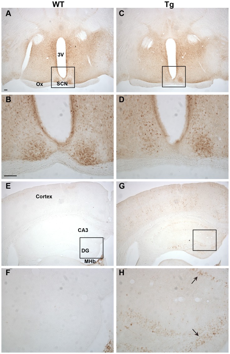
Figure 5. Ngb-IR in the hypothalamus and hippocampus.
No difference could be seen in localization or intensity of Ngb-IR in the hypothalamus of WT (A-B) and Tg mice (C-D). The hippocampus was voided of Ngb-IR in WT mice (E-F). In Tg mice Ngb-IR was seen in cell bodies (black arrow) and processes of most structures of the hippocampus (G-H). Abbreviations: 3rd ventricle (3V); field CA3 of hippocampus (CA3); Dentate gyrus (DG); Medial habenular nucleus (MHb); Optic chiasm (Ox). Scale bar 100 µm.

