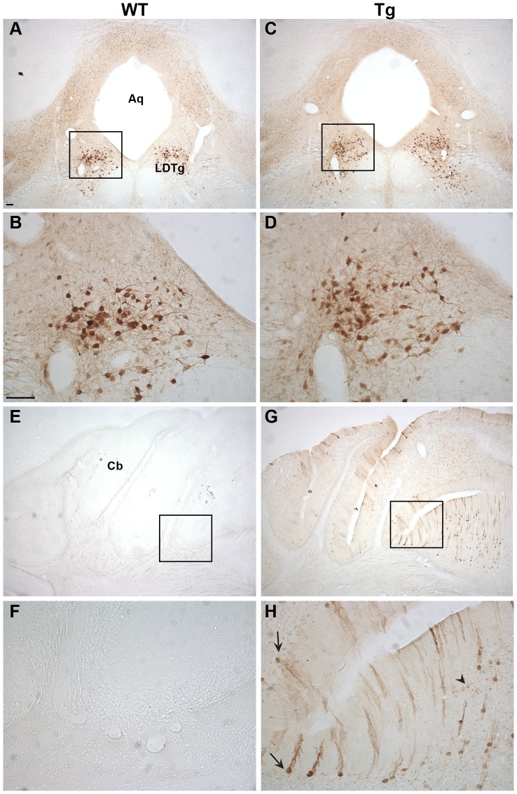
Figure 6. Ngb-IR in the hindbrain and cerebellum.
In the hindbrain pontine nuclei no difference was seen between WT (A-B) and Tg (C-D) mice in localization, intensity or morphology of the Ngb-IR neurons. No Ngb-IR could be seen in the cerebellum of WT mice (E-F). In contrast, Ngb-IR was seen in Purkinje cells (black arrow) and granule cells (black arrowhead) of the cerebellar lobule in Tg mice (G-H). Abbreviations: Aqueduct (Aq); Cerebellum (Cb); Laterodorsal tegmental nucleus (LDTg). Scale bar 100 µm.

