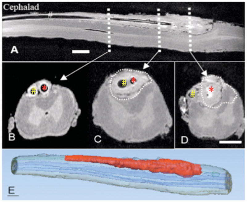
Figure 2.

Ex Vivo MRI (magnetic resonance imaging) taken post mortem through the lumbar spinal cord of the dog with sampling catheter (red arrow) and infusion catheter (white arrow) that received a 28-day infusion of intrathecal morphine sulfate (12 mg/d). A: Mid-sagittal plane through the lumbar cord. B,C,D: shows three transverse sections taken at the levels indicated. In Figure B, sampling catheter and infusion catheter indicated by red and white arrows, respectively. Size bars are approximately 0.3 cm. The C and D sections correspond to the levels in the longitudinal section that are proximal to the respective catheter tip. E: Three dimensional reconstruction of dog spinal cord from ex vivo MRI. Red and blue volumes indicate granulomatous mass and spinal cord, respectively.
