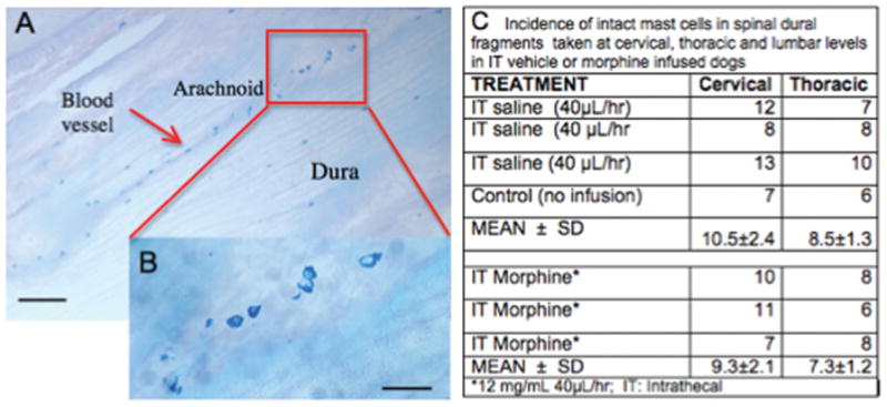
Figure 4.

A: Section showing Alcian Blue stained lumbar dura mast cells in control animal at low (bar = 100μ). B. Inset showing enlargement of marked area at high (bar = 30μ) power. Note alignment of cells in arachnoid with blood vessel. C: Table presenting counts of intact Alcian Blue staining profiles in cervical, thoracic and lumbar dura in three animals receiving intrathecal (IT) saline or 3 animals receiving IT Morphine (12mg/d).
