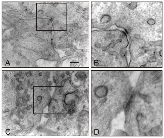
Figure 7. EW induces junctional contact formation in TCam-2 cells.
Transmission electron microscopy pictures showing EW induced TCam-2 cell-cell junctional contacts. In A one typical cell-cell adhesion junction is showed and in B an higher magnification of the same junction is reported. In C cellular contact compatible with gap-junction structure is shown and in D a higher magnification of the same junction is reported. Bar, A and C 226 nm B and D: 150 nm.

