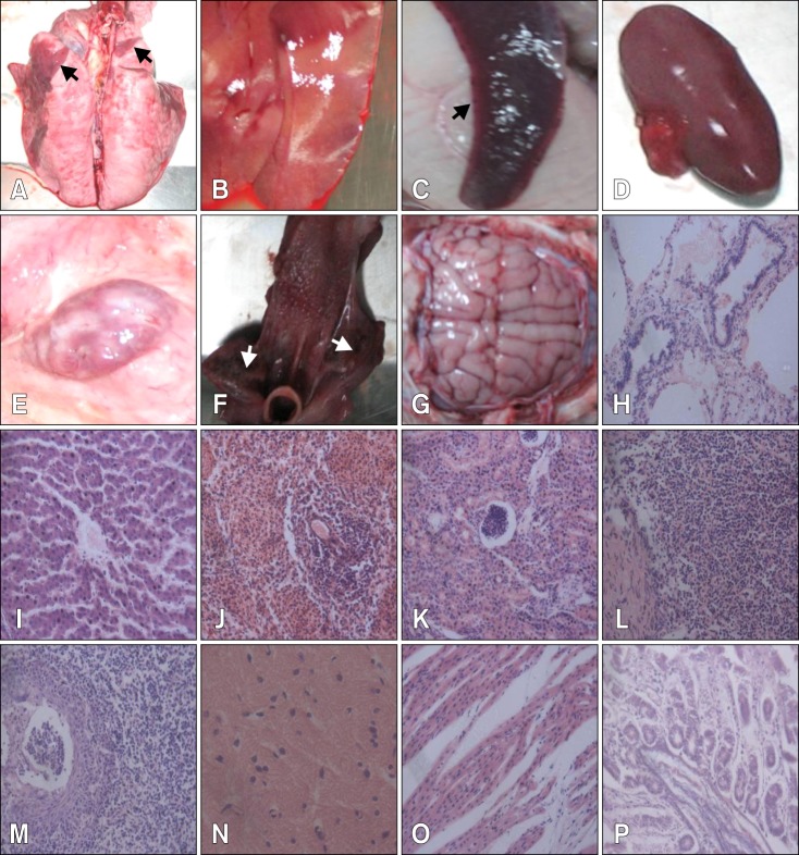
Fig. 2.
Severe damage to multiple organs in experimental, infected piglets by postmortem and histopathological examinations. (A) Lung necrosis (arrows). (B) Liver with yellowish white spots indicating necrosis or hemorrhage. (C) Spleen infarct (arrow). (D) Kidney with bleeding spots. (E) Hemorrhagic lymph node. (F) Tonsil necrosis (arrows). (G) Slight encephalic edema. (H) Alveolar ducts and terminal bronchiolar cavities filled with cellular and serous exudates. (I) Swelling and degeneration of liver cells. (J) Splenic cord with unclear structure and reduced lymphocytes. (K) Swelling and disintegration of epithelial cells. (L) Reduced lymphoid nodules with irregular structures. (M) Epithelial cells filled with eosinophilic intranuclear inclusions. (N) Glial cells with neurons. (O) Breakage and disintegration of myocardial fibers. (P) Midgut gland atrophy. H&E stain, ×400.

