
Figure 3. A cell intrinsic role for miR-155 in promoting effector CD8+T cells.
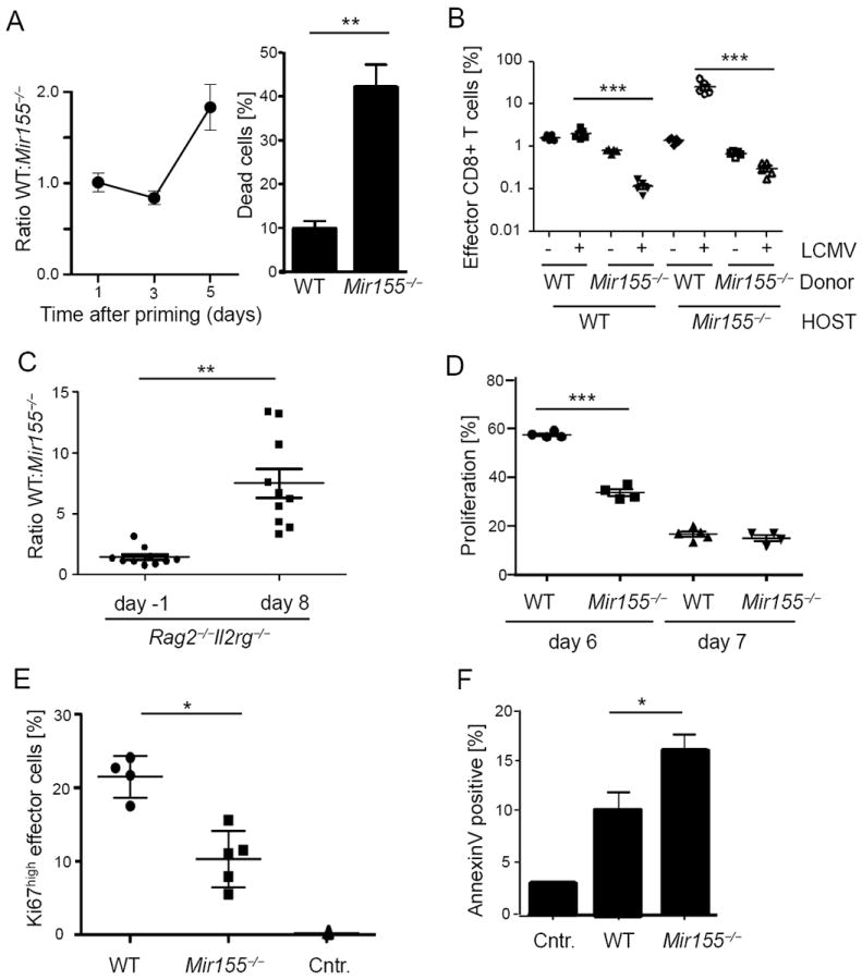
(A) Congenically marked wild type (WT) and Mir155-/- OT-1 cells were competitively cocultured with peptide pulsed dendritic cells and the ratio of populations is pictured at indicated time points (left). On day 5, percentage of trypan blue cells harvested from either WT or Mir155-/- cultures was counted (right graph). Pooled data from three representative experiments are pictured. (B) CD8+ T cells from WT and Mir155-/- mice were cotransferred into WT or deficient hosts before LCMV WE infection and percentages in blood at day 8 were measured. (C) A 1:1 mix of WT and Mir155-/- splenocytes was adoptively transferred into Rag2 and IL2Rγ double deficient mice which were infected with LCMV WE two months after transfer. CD8+ effector T cell ratios at days -1 and 8 post infection are pictured. (D) Proliferating BrdU positive splenic CD44high CD8+ effector T cells at days 6 and 7 post infection. (E) At the same time, cells were stained for the proliferation marker Ki67 (day7) and (F) apoptotic cells were identified by AnnexinV staining. Symbols represent individual mice, and the line is the mean +/- SEM. Representative results from two (B, C) to three (D-F) experiments are pictured.