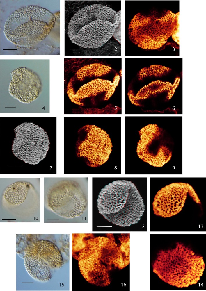
Plate I.
Scale bars: 10 μm. (1), Pollen Type I, specimen A, LM image (high focus); (2), Pollen Type I, specimen A, CLSM image, total stack image/projection, anaglyph; (3), Pollen Type I, specimen A, CLSM partial stack image, proximal side; (4), Pollen Type I, specimen B, LM image (median focus); (5), Pollen Type I, specimen A, CLSM partial stack image, distal side; (6), Pollen Type I, specimen A, CLSM single image, optical section; (7), Pollen Type I, specimen B, CLSM total stack image/projection, anaglyph; (8), Pollen Type I, specimen B, CLSM partial stack image, lateral view (high focus); (9), Pollen Type I, specimen B, CLSM partial stack image, lateral view (low focus); (10), Pollen Type II, specimen A, LM image, high focus; (11), Pollen Type II, specimen A, LM image, low focus; (12), Pollen Type II, specimen A, CLSM total stack image/projection, anaglyph; (13), Pollen Type II, specimen A, CLSM partial stack image, lateral view on distal side (high focus); (14), Pollen Type II, specimen A, CLSM partial stack image, lateral view on proximal side (low focus); (15), Pollen Type II, specimen B, LM image, high focus; (16), Pollen Type II, specimen B, CLSM total stack image/projection.

