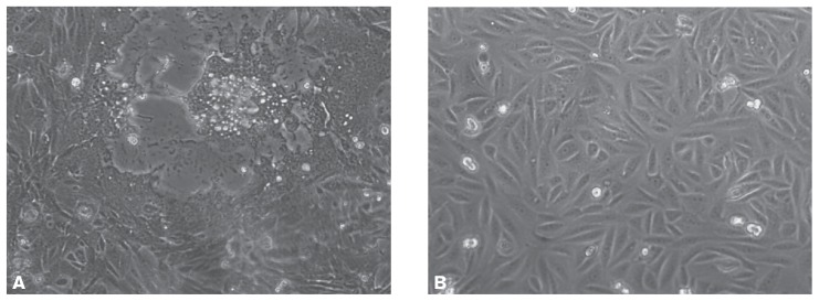
Figure 1.
Vero cells grown for 24 h and then inoculated (A) with canine distemper virus (CDV) or not inoculated (B). After 72 h about 90% of the CDV-inoculated Vero cells showed cytopathic effects. Images were acquired with a Leica DM IRB microscope (Leica Microsystems, Heerbrugg, Germany) at a magnification of × 200.

