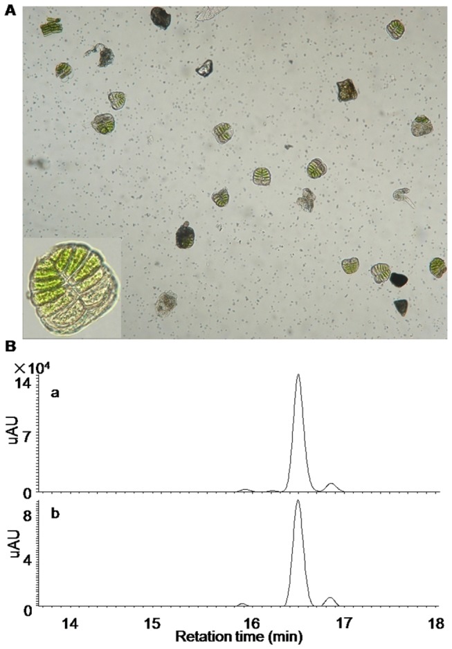
Figure 4. LC-MS analysis of the extracts from the isolated glandular trichomes of X. strumarium.

(A) The glandular cells purified from Hubei-Wuhan X. strumarium species (inset: a single glandular structure at a magnified visual field); (B) the comparison of the LC-MS profiles derived from the chloroform-dipping extracts (a) and the extracts directed from the isolated glandular trichomes (b).
