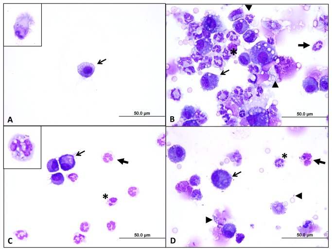
Figure 6. Cytologic evaluation of BALF from infected mice.
Bronchoalveolar lavage fluids from control and infected animals (see Figure 5) were concentrated onto a glass slide, fixed with methanol, air-dried, stained with modified Wright-Giemsa, and examined by microscopy (100X objective). Panel A shows a representative field of a sample collected from a mouse inoculated with sterile PBS (control). These control samples are low in cellularity and consist of occasional macrophages (arrow) and ciliated, columnar respiratory epithelial cells (inset). Panel B shows a representative field of a sample from a mouse infected with 104 B. mallei bacteria. Large numbers of degenerate neutrophils (block arrows) and foamy macrophages (arrows) are seen admixed with cellular debris and occasional red blood cells. Medium-sized bacilli are seen phagocytosed by neutrophils (asterisk). Extracellular bacteria are also present (arrowheads). Panel C is a representative field of a sample from mouse infected with 103 B. mallei cells, and Panel D is a representative field of a sample from mouse infected with 103 B. pseudomallei CFU. In both panels, moderate numbers of degenerate neutrophils and foamy macrophages are present, along with intracellular bacilli, illustrating the similar type and magnitude of inflammation elicited by the two species at the same dose. The inset in Panel C demonstrates that neutrophils contain bacilli adhered to their surface and within the lumens of intracytoplasmic vacuoles.

