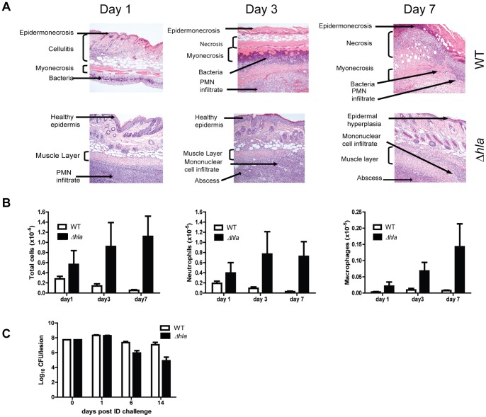
Figure 2. AT is a major contributor to disease severity in S. aureus dermonecrosis.
BALB/c mice (n = 10) were infected ID with 5×10⁷ cfu WT (□) or Δhla (▪). (A) Representative H&E stained histological sections 1, 3 and 7 days post-infection. (B) BALB/c mice (n = 5 for days1 and3, n = 10 for day 7) were infected ID with WT or Δhla. Skin lesions were collected 1, 3 or 7 days post-infection and the neutrophils and macrophages were enumerated by flow cytometry as described in the methods. Cell number differences between WT and Δhla infected mice were calculated with a Student’s t-test, and considered statistically difference if p<0.05 (indicated as*). (C) Bacterial CFU in skin lesions. Skins were harvested, homogenized for bacterial enumeration 1, 6, 9 and 14 days post infection (5 mice for days 1 and 6, 10 mice for days 9 and 14). Data were analyzed using a Mann-Whitney U test, and were statically significant after day 6 (p≤0.002).

