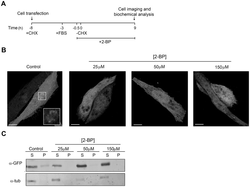
Figure 1. 2-BP inhibits acylation and membrane association of newly synthesized N13GAP-43(C3S).
A) Schematic representation of the experimental procedure used in B and C. The CHO-K1 cells were transfected at −8 h with the plasmid encoding N13GAP-43(C3S)-YFP. At 30 min before CHX withdrawal (-CHX), cells were incubated with 25, 50 or 150 µM 2-BP or DMSO (vehicle, Control). At 0 h, CHX was removed and cells were further incubated with 2-BP at the concentrations indicated above, or with DMSO, at 37°C for 9 h. Finally, cells were analyzed by confocal fluorescent microscopy or subjected to biochemical assays. B) Representative images showing the effect of 2-BP or DMSO (Control) on the TGN association of N13GAP-43(C3S). The fluorescent signal from YFP was pseudocoloured gray. The inset shows details of the boxed area at a higher magnification. Scale bars: 5 µm. C) After treatment with 2-BP, CHO-K1 cells transiently expressing N13GAP-43(C3S)-YFP were lysed, ultracentrifuged and the supernatant (S) and pellet (P) fractions were recovered. Proteins from these fractions were western blotted with an antibody to GFP (α-GFP) and α-tubulin (α-tub).

