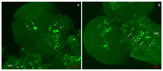
Figure 4. Neurons in the larval CNS stained with CCHamide-2 antisera.
These preparations are somewhat compressed, because of mounting without the application of spacer rings. These spacer rings were present in the preparations shown in Figure 6 and Figure 7. For a schematic representation of the neurons and neuropil in the two larval brain hemispheres and ventral nerve cord, see Figure 5. A. Ventral view of one hemisphere of the brain, showing 7-8 immunoreactive neurons located in the central region (long arrows) and processes (neuropil) projecting to other regions of the brain (short arrow). This neuropil corresponds to the neuropil indicated by number 3 in Figure 5. A piece of the ring gland (asterisk) is also visible, as well as a piece of the ventral nerve cord (VNC). Scale bar = 50 µm. B. Ventral view of another brain hemisphere with the ventral nerve cord. At least 12 nerve cells (short arrows) can be seen here symmetrically ordered along the midline of the ventral nerve cord (VNC). Scale bar = 50 µm.

