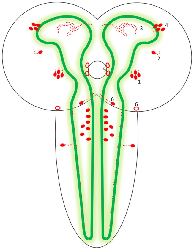
Figure 5. Schematic drawing of the localizations of the CCHamide-2 immunoreactive neurons and neuropil in the CNS of third instar larvae.

The drawing shows the two hemispheres and the ventral nerve cord. The central neuropil of the larval CNS (stained with a synapsin mouse monoclonal antibody, cf. Figure 6 and Figure 7) is outlined by green lines and shades. Neuronal perikarya and neuropil are drawn in red. Weakly immunoreactive perikarya are drawn as open red symbols. The perikarya indicated by 1 and 2 in the right hemisphere are located dorsally in each hemisphere; the perikarya indicated by 4, and 5, and 6 are located in the ventral parts of the hemispheres. The neuropils indicated by 3 as red dots in the anterior parts of the central neuropil are located partially ventrally and partially medially between the levels of the neurons 1 and 4. All perikarya in the ventral nerve cord are located in the ventral part of this nerve cord. They belong to the three fused thoracic ganglia.
