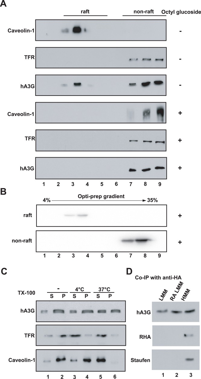
Figure 2. Steady state hA3G in the cytoplasm appears in three different forms. A.
293T cells expressing HA tagged hA3G were lysed in hypotonic TE buffer. As described in Methods, P100 was prepared and treated with or without nonionic detergent octyl glucoside as indicated, then resolved by the sucrose floatation assay into the raft and non-raft proteins. Each fraction was analyzed by Western blot for the presence of hA3G, Caveolin-1 and TFR. B. The raft and non-raft fractions of hA3G were collected and treated with octyl glucoside, then resolved in the Opti-prep velocity gradient. Western blots of each fraction were probed with anti-HA. C. 293T cells expressing HA tagged hA3G were lysed in hypotonic TE buffer, and the S1 fractions were either untreated (lane 1 and 2) or treated with 0.5% Triton X-100 at 4°C (lane 3 and 4) and 37°C (lane 5 and 6), respectively. Following the ultra-centrifugation of the S1 fraction, Western blots of the P100 and S100 fractions were then probed with antibody specific for HA (top), TFR (middle), and caveolin-1 (bottom). S and P represent the S100 and P100 fractions, respectively. D. Fractions that respectively contain the soluble LMM (lane 1), RA LMM (lane 2) and HMM (lane 3), were subjected to immunoprecipitation with anti-HA, followed by Western blots of the immunoprecipitates probed with anti-HA, anti-RHA, and anti-Staufen, respectively.

