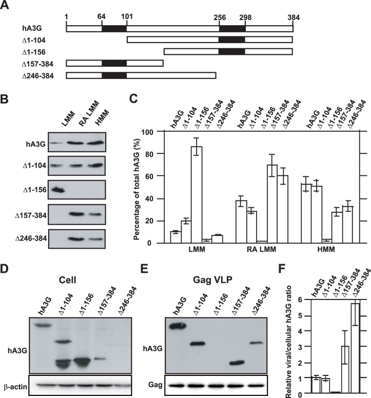
Figure 4. The correlation between the cellular distribution and viral incorporation of hA3G. A.
Wild type and mutant hA3G. The filled rectangles represent the two zinc coordination units. The numbers represent the amino acid positions. B. 293T cells were co-transfected with hGag and either wild-type or mutated forms of hA3G, and the S1 fractions of the cell lysates were subjected to ultra-centrifugation and octyl glucoside treatment, resulting in the LMM, RA LMM and HMM forms of hA3G. The amounts of hA3G in the three forms were determined by Western blot. C. The cellular distributions of wild type and mutant hA3G are graphically shown. D. Western blot of cell lysates were probed with anti-HA (top) and anti-β-action (bottom). E. Western blot of virus like particle lysates probed with anti-HA (top) and anti-p24 (bottom). F. The relative amounts of mutated hA3G in the cell or viral lysates were normalized to wild-type hA3G, and then a ratio of viral to cellular hA3G was determined and used to measure its ability to be packaged into virions. The bar graphs in panel C and F represent the means of results of experiments performed at least three times, and the error bars represent standard deviations.

