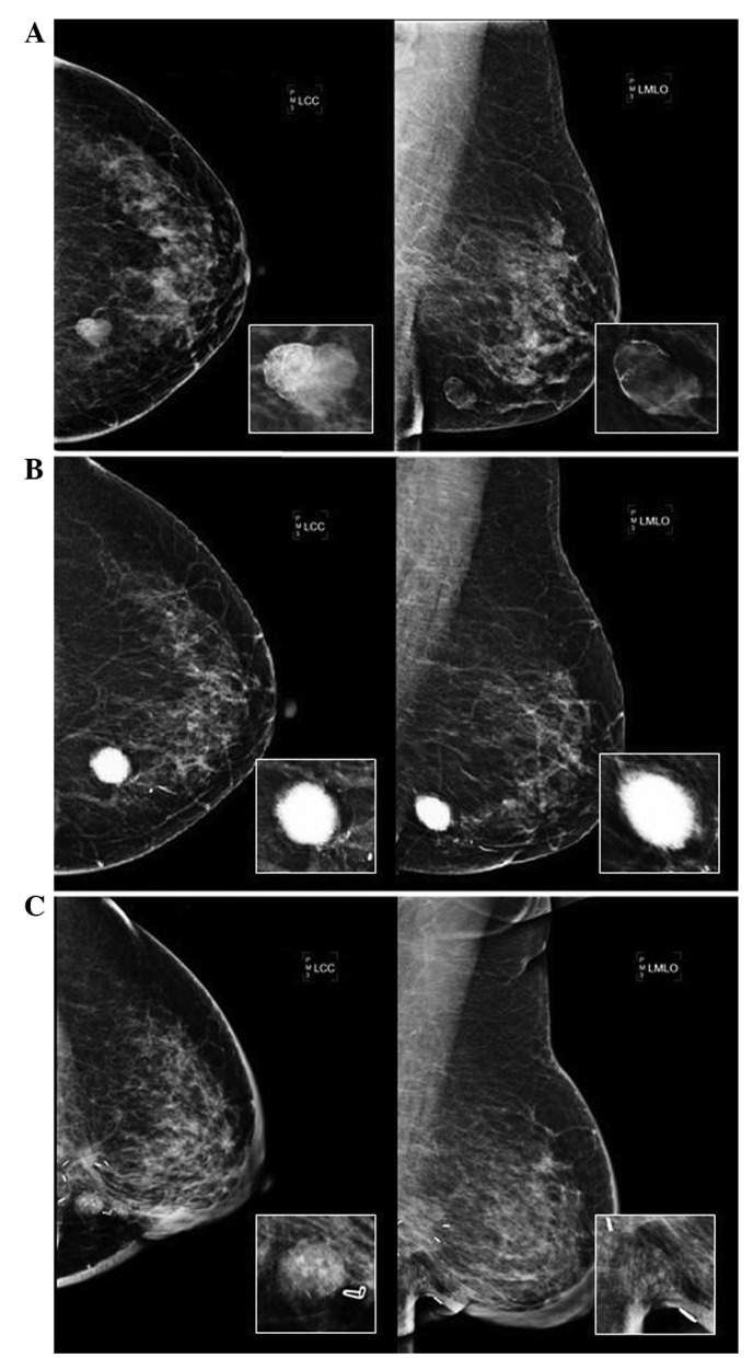
Figure 1.

(A) Mammography performed in 2005. CC and MLO views show an oval, well-circumscribed mass, isodensity with a rim and calcification. (B) Mammography performed in 2009. CC and MLO views show a large calcified mass at the left inferior inner quadrant. The magnified pictures reveal the sunburst appearance, which resembled osteosarcoma calcification. (C) Mammography performed in 2010. CC and MLO views show a well-defined 1.5-cm mass with multiple coarse calcifications underneath the surgical scar. CC, cranio-caudal; MLO, medio-lateral oblique.
