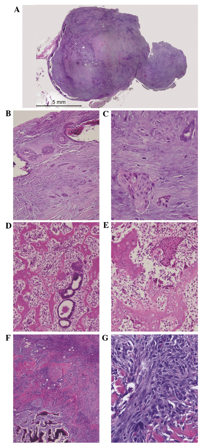
Figure 2.

(A–C) First mass removed in 2005. (A) Low-grade adenosquamous carcinoma with focal osseous metaplasia, initially diagnosed as nonmalignant fibroadenomatous nodule (H&E). (B) Presence of banal-appearing epithelial component in fibromyxoid stroma (H&E; magnification, ×200). (C) Low-grade angulated tubules and irregular squamous nests (H&E; magnification, ×400). (D and E) Recurrent tumor in 2009. (D) Prominent osteosarcomatous component with few entrapped glands (H&E; magnification, ×200). (E) Entrapped squamous epithelium (H&E; magnification, ×200). (F and G) Recurrent tumor in 2010. (F) Focal osseous component (H&E; magnification, ×40). (G) Predominant spindle cell morphology without any epithelial cells documented (H&E; magnification, ×400). H&E, hematoxylin and eosin.
