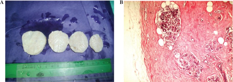
Figure 2.
Gross and microscopic appearence of fibroadenolipoma. (A) Lesion is a well-circumscribed, encapsulated, greyish-white-colored mass resembling fibroadenoma. Note the small cystic spaces on the cut surface. (B) Hematoxylin and eosin stained mammary glandular tissue with mature adipocytes in hyalinized fibrous tissue (magnification, ×40).

