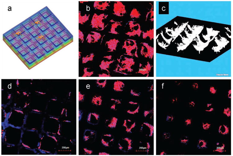
Figure 5.
Culture of 3T3 Cells on the 20% PEGDA Stepwise Microwells. a) Schematic view of the cell culture on stepwise microwells. b) Confocal z-projection image of 3T3 cell culture after 4 days illustrated the microwells scaffold can support cell adhesion and proliferation. c) 3D reconstruction of 3T3 cells on the microwells scaffold via Image J displayed bowl-like multicellular organizations inside the pocket area of the wells, which indicated the geometric guidance of the scaffolds. d–f) The z-sectioning images from the top to the bottom of the scaffold, showing different cell-cell organizations corresponding to the altering scaffold geometry at different z positions. F-actin (red) was stained with rhodamine phalloidin and the nuclei (blue) were counterstained with Hoechst.

