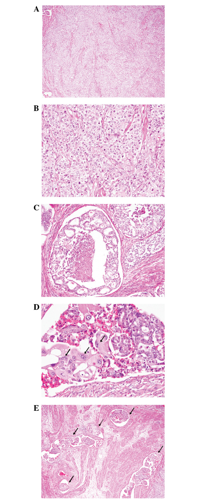
Figure 2.

Histopathological findings of the uterine corpus tumor. (A) Poorly-differentiated endometrioid adenocarcinoma component. Proliferation of sheets or variable-sized nests of atypical epithelial cells. (hematoxylin and eosin; magnification, ×40). (B) Atypical epithelial cells contain a relatively rich, marginally eosinophilic cytoplasm and large nuclei with coarse chromatin and small nucleoli (hematoxylin and eosin; magnification, ×200). (C) Glandular differentiation showing a cribriform structure with central necrosis (hematoxylin and eosin; magnification ×100). (D) Choriocarcinomatous component. Syncytial giant cells are scattered (arrows; hematoxylin and eosin; magnification, ×200). (E) Vascular and lymphatic invasions are prominent (arrows; hematoxylin and eosin; magnification, ×40).
