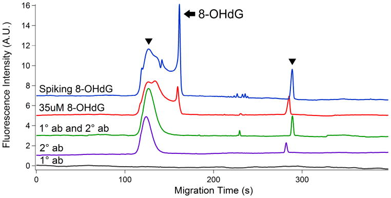
Figure 2.

Electropherograms of validation tests in positive and negative controls. (1) positive control #1-- a complex with 3530 uM 8-OHdG standard; (2) Positive control #2 -- a complex with 35 uM 8-OHdG standard; (3) Negative control #1-- a complex with primary and secondary antibody; (4) Negative control #2 -- secondary antibody alone; (5) Negative control #3 -- primary antibody alone. Hydrodynamic injection at pH 9.5 and 3.4 kPa for 5 s; separation in 20 mM sodium tetraborate buffer at 17 kV. Arrowhead: secondary antibody; Arrow: 8-OHdG. Top trace is offset in the y-axis for the clarity.
