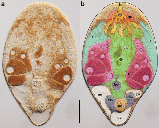
Fig. 3.

Image of a mature and live specimen of Isodiametra pulchra without (left) and with superimposed colors (right) to illustrate the general morphology of acoels. From top to bottom: yellow: frontal organ (fo); red: nervous system (ns); green: central syncytium (cs); cyan: testes (t); pink: ovaries (o); gray: mouth; purple: female copulatory organs (fco) composed of seminal bursa, bursal nozzle, and vestibulum (from posterior to anterior); white: chordoid vacuoles (cv); blue: false seminal vesicles and prostatoid glands (fsv); orange: male copulatory organ (cop) composed of muscular seminal vesicle and invaginated penis. Scale bar: 100 μm
