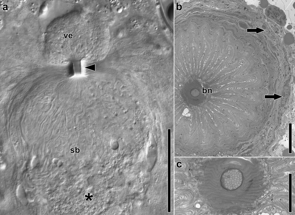
Fig. 6.

Female copulatory organs in Isodiametra pulchra. a Image of female copulatory organs in a live and squeezed specimen. Note the mass of elongated and convoluted sperm in the seminal bursa (sb) that merge towards the bursal nozzle (arrowhead) and a few “heads” extending into the vestibulum (ve). Asterisk marks bursal stalk connecting the bursa with the digestive parenchyma, arrowhead points to bursal nozzle. b Electron micrograph showing cross section through the bursal nozzle (bn). Arrows point to nuclei of cells of the bursal wall. c Counterclockwise rotated detail of b. Note the density of sperm in the duct of the bursal nozzle. Abbreviations: bn bursal nozzle; sb seminal bursa; ve vestibulum. Scale bars: a 50 μm; b 10 μm; c 5 μm
