Figure 3.
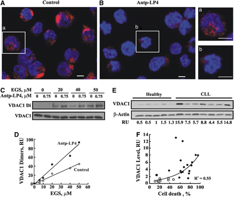
Antp-LP4 induces HK displacement and VDAC1oligomerization. MEC-1 cells were incubated without (control, A) or with Antp-LP4 (0.5 μM, 90 min, B) and HK detachment from the mitochondria was analyzed by immunostaining using anti-HK-I antibodies. Representative microscopic fields from one of three similar experiments are shown. (a) and (b) show an enlargement of a representative cell from A and B, respectively. Scale bar, 5 μm. To assess VDAC1 oligomerization, control and Antp-LP4-treated (0.75 μM, 40 min) MEC-1 cells were incubated with the indicated concentration of EGS (15 min, 30°C). Cross-linking was terminated by sample buffer addition and heating (70°C, 10 min). Cells were subjected to SDS-PAGE, followed by immunoblotting using anti-VDAC1 antibodies and HRP-conjugated secondary antibody, visualized by enhanced chemiluminescence (C). Quantitation of immuno-stained VDAC1dimers as a function of EGS concentration (D). VDAC1 over-expression in CLL patients-derived PBMCs and healthy donors as probed with anti-VDAC1and anti-β-actin antibodies. A representative immunoblot and its quantitation (RU) are shown (E). VDAC1 levels in PBMCs isolated from healthy donors (n=9) (o) or CLL patients (n=18) (●) were plotted as a function of the percentage of cell death induced by Antp-LP4 (1.5 μM) for each individual (R2=0.55) (F)

