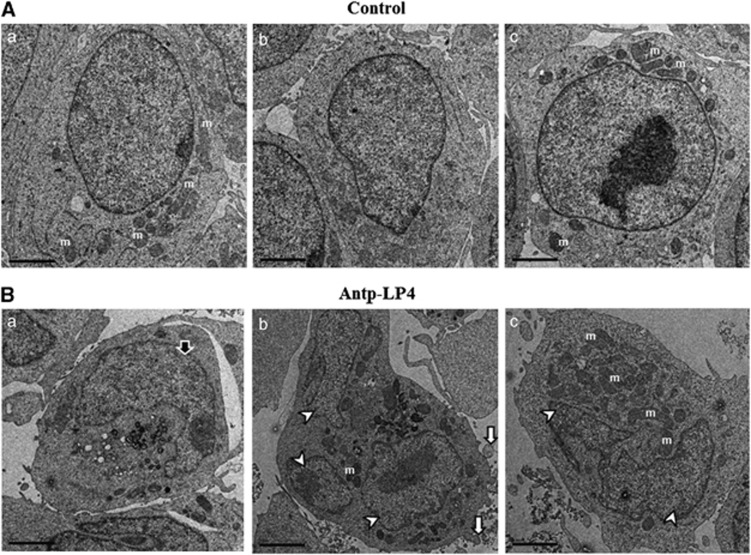
Figure 5.
Electron microscopy visualization of Antp-LP4 induces apoptotic cell death MEC-1 cells left untreated (A) (control) or treated with 1.5 μM of Antp-LP4 (B) were examined by EM. Cells from three representative control and peptide-treated samples are presented. In the peptide-treated cells, typical morphologic changes associated with apoptosis, such as condensation of nuclei (Ba, black arrow), DNA fragmentation (Bb, Bc, white arrowheads) and membrane blebbeing (Bb, white arrow), were noted. Mitochondrion is indicated by letter m

