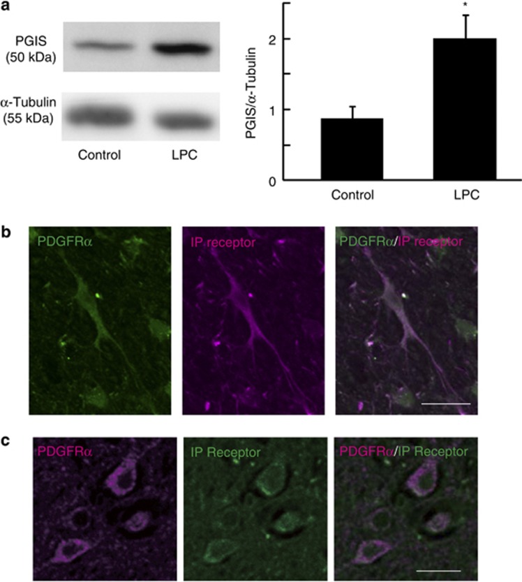
Figure 3.
Prostacyclin and its receptor are expressed in the CNS tissue of mouse and MS patients. (a) Representative spinal cord sections immunostained for IP receptor (magenta) and PDGFRα (green) in control mice. Scale bar, 20 μm. (b) Representative brain section immunostained for IP receptor (green) and PDGFRα (magenta) in MS patients. Scale bar, 10 μm. (c) Western blot analyses for PGIS and α-tubulin expression in the spinal cord. Spinal cord tissues were obtained 4 days after LPC injection. PGIS level was normalized to that of α-tubulin. Values represent the mean±S.E.M. (n=6). *P<0.05 compared with control

